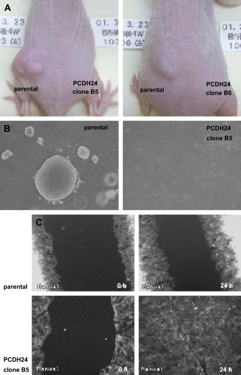
Figure 1.

Influence of PCDH24 expression on growth of HCT116 cells. (A) Representative in vivo growth of parental and PCDH24‐expressing HCT116 cells. Each type of cell (3×10⁶cells) was inoculated into the right and left back of the same athymic BALB/cA Jcl‐nu/nu mice (6–7 weeks old, male; CLEA Japan, Inc., Tokyo), and photographs were taken after 4 weeks. The tumor size of parental cells was calculated according to the formula: length (mm)×width (mm)×height (mm)×0.5236 was 206.3±135.9cm3 (n=4). All procedures were approved by the committee on animal care. (B) Representative image of soft agar assays. A total of 3×10²cells were plated and the colonies in triplicate wells were counted after 11 days. Data are represented as mean±SD: parental cells (297±18.3), PCDH24‐expression clone B5 (0±0). (C) Effect on cell motility. Photographs were taken before (0h) and 24h (24h) following wound injury.
