
Figure 1.
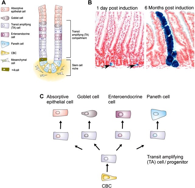
Anatomy of the intestine. A) Schematic representation of the crypt‐villus axis. B) Lineage tracing experiments demonstrating that LGR5 cells are the stem cells of the intestine. Blue ribbons show the expression of LacZ from the Rosa26LacZ locus upon induction of CRE enzyme with tamoxifen, on the LGR5EGFP‐IRES‐CREert/Rosa26R mouse. After one day single CBC cells are induced. Six months later, entire blue ribbons indicate that the CBC cells that had an activated Rosa26LacZ are still producing all the cells of the intestinal epithelium. C) Differentiation hierarchy of the small intestine.