Figure 3.

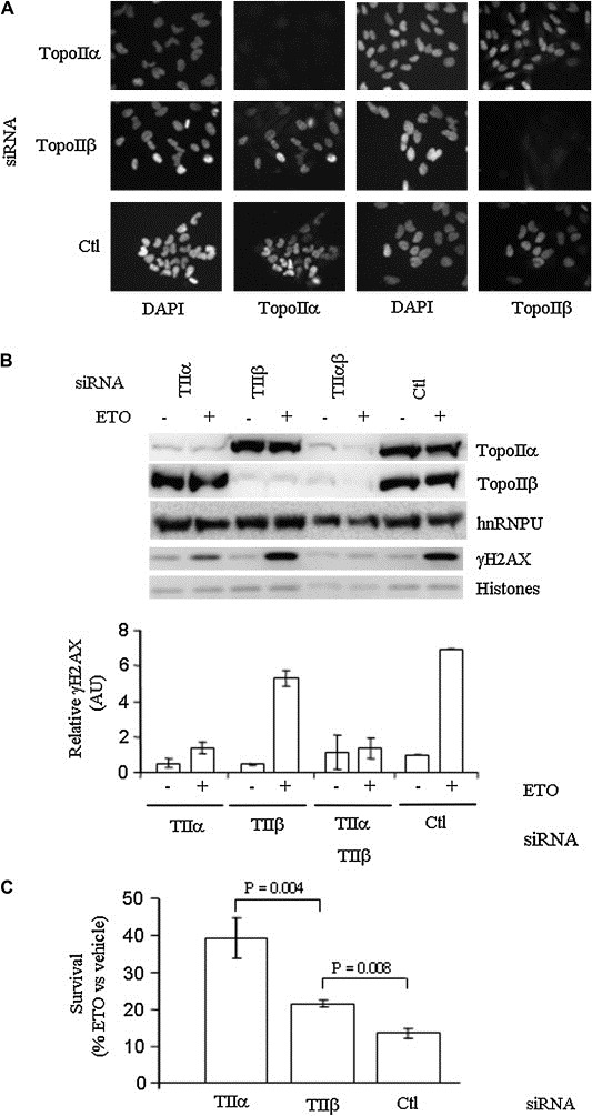
Etoposide cytotoxicity and induction of γH2AX in HeLa cells depends on topoIIα. (A) Indirect immunofluorescence of topoIIα and topoIIβ following siRNA transfection compared with DAPI nuclear staining. Ctl = siRNA directed against GFP. (B) Top, Western blot of γH2AX, topoIIα and topoIIβ subsequent to siRNA and etoposide treatment, compared with hnRNPU and total histone levels (Ponceau stain). Bottom, γH2AX levels (mean ± SD). Quantifications were normalized to either hnRNPU (topoII) or total histones (γH2AX). (C) HeLa cell survival 6 days following etoposide treatment of Ctl and topoII siRNA transfected cells analyzed by formazan formation assay. Values are ±SD of three independent experiments performed in duplicate.
