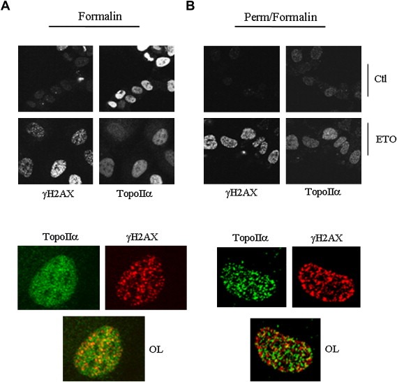
Figure 4.

TopoIIα and γH2AX display largely distinct nuclear distributions with some signal overlap. HeLa cells were treated with 25μM etoposide for 1h, were either directly fixed in formalin. (A) or pre‐permeabilized with 25mM Hepes, pH 7.5, 0.3M NaCl, 1.5mM MgCl2, 0.2mM EDTA, 0.5% Triton X‐100, 0.5mM DTT and 1mM phenylmethylsulfonyl fluoride for 2min prior to formalin fixation. (B) Coverslips were then processed for immunocytochemistry using the indicated antibodies. Top, wide field view; bottom, magnification of cells from the 1h etoposide panel; OL, overlay of the two signals. Representative cells are shown. Similar results were obtained following 10min and 20min treatments.
