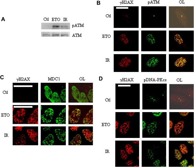
Figure 6.

γH2AX foci mark sites of DNA repair activity. (A) ATM and pATM levels in HeLa cells treated with vehicle (Ctl), 25μM etoposide (1h) or 6Gy IR (IR). (B) Co‐localization (yellow signal in the overlay (OL)) of γH2AX (red) and pATM (green). Treatment with vehicle (Ctl), IR and etoposide (ETO) are as indicated. Scale bar=20μm. (C,D) Co‐localization of γH2AX with MDC1 (D), and pDNA‐PK (E). In B and E, the Ctl overlays were intensified in order to visualize the cells.
