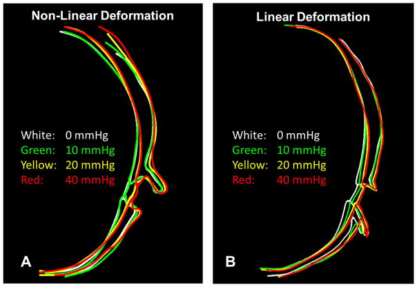
Figure 7.
Delineations of the posterior sclera and optic nerve head demonstrate scleral bowing and canal expansion with increasing IOP. To notice more clearly the outward bowing of the sclera and widening of the canal, the outlines were registered to one another in the peripheral sclera (notice the point where all lines cross) A. In one eye a non-linear deformation pattern was observed. Between 0 and 10 mmHg, little change was seen in the posterior sclera as the globe returned to its physiological shape. Between 10 and 20 mmHg the peripapillary sclera bowed outward and the lamina began to cup. Between 20 and 40 mmHg the sclera underwent only a small displacement, but noticeable deformation was seen in the canal and lamina surface. The delineations for this eye are shown on the original images in Supplementary Figure 2B. In a second eye, the deformation pattern was much more linear with moderate scleral bowing observed at each pressure change and little deformation of the lamina cribrosa surface. The delineations for this eye are shown on the original images in Supplementary Figure 3.

