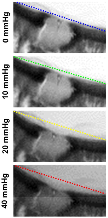
Figure 8.
Detailed view of axial cross sections through the optic nerve head and peripapillary sclera showing outward bowing of the sclera with increased IOP. Straight lines connecting the most distal sclera in the images are overlaid to help notice the deformations. Note also the detail and substantial deformations of structures posterior to the sclera, typically hidden from OCT due to shadowing.

