Figure 9.
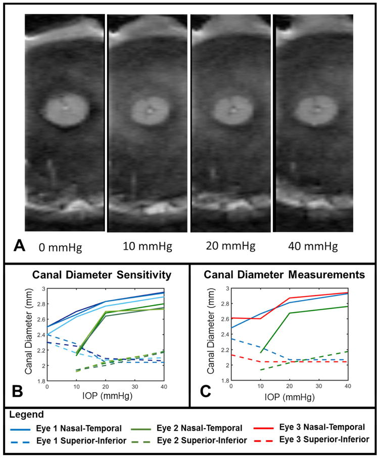
The scleral canal underwent measurable expansion due to IOP. A. Coronal views showing the scleral canal under 0, 10, 20 and 40 mmHg. The superior-inferior axis is shown top to bottom. B. Measurements of the canal diameters were marked in triplicate to determine measurement sensitivity and the average standard deviation of the triplicate measurements across all eyes, pressures, and axes was 30 μm. C. The nasal-temporal scleral canal diameter (solid lines) increased measurably due to IOP changes. The increase was greatest between IOPs of 10 and 20 mmHg. The superior-inferior canal diameter (dashed lines) only changed measurably in eye 2 (Green dashed line between 10 and 20 mmHg). In eye 2 we could not confidently identify a section at 0 mmHg that could be compared to the other pressures conditions and this condition was not considered in our measurements. Note that the resolution of this scan is higher in the nasal-temporal direction (left-right) than in the superior-inferior direction (top-bottom). Recall that sheep eyes have a scleral canal that is typically more elongated in the nasal-temporal direction than in the superior-inferior one.

