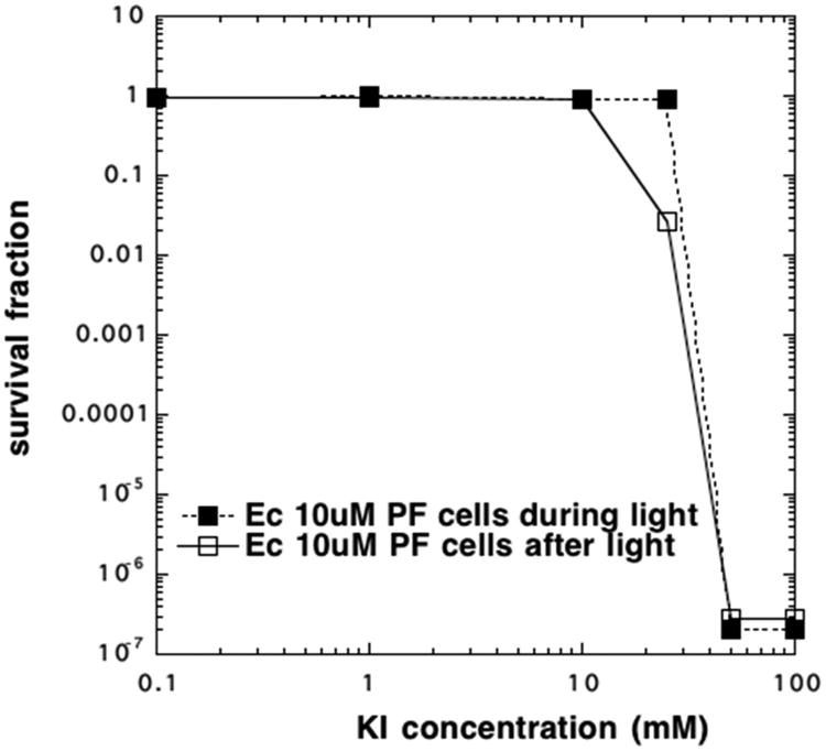
Figure 2.

Comparison of aPDI killing with cells present and cells added after light. PF (10 μM) was exposed to 10 J/cm2 blue light in the presence of different concentrations of KI. E. coli cells (108/mL) were either present during light or added after light.
