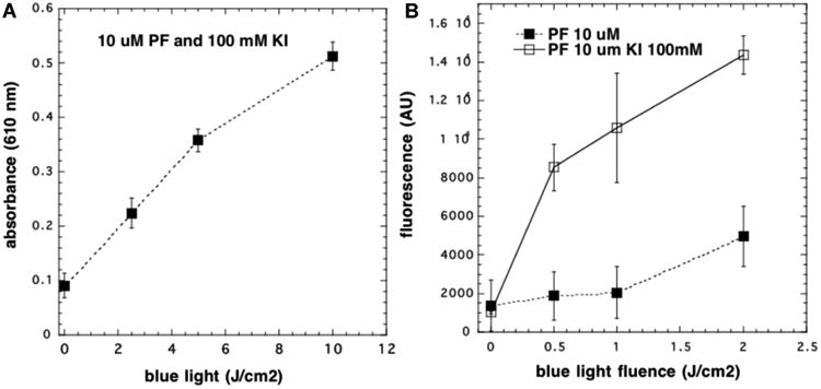
Figure 5.

Formation of starch–iodine and hydrogen peroxide in solution (no cells): (A) PF (10 μM) and KI (100 mM) were illuminated with increasing fluence of 415 nm light and aliquots withdrawn and added to starch solution; (B) same as (A) but included PF without KI, and aliquots were withdrawn and added to Amplex Red reagent.
