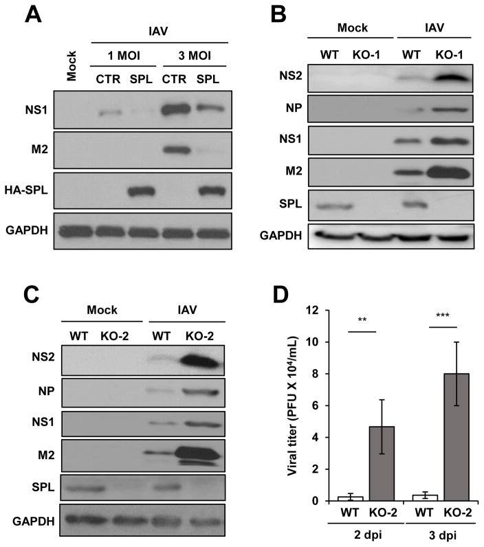
Figure 1. SPL inhibits the replication of influenza virus.
(A) A549 cells (2 X 10⁵) were transfected with empty vector control DNA (CTR) or HA-tagged SPL (HA-SPL)-encoding DNA. One day later, cells were either mock-infected (Mock) or infected with 1 or 3 multiplicity of infection (MOI) of IAV. At 8 hr post-infection (hpi), cells were harvested for Western blot analysis to check the level of NS1, M2, HA-SPL, and GAPDH. (B and C) WT, KO-1 cells (B), and KO-2 cells (1 X 10⁶) (C) were infected with 0.1 MOI of IAV or mock-infected. At 1 day post–infection (dpi), Western blotting was performed to detect NS2, NP, NS1, M2, SPL, and GAPDH proteins. (D) WT or KO-2 cells (2 X 10⁵) were infected with 0.01 MOI of IAV. At 2dpi and 3dpi, plaque assay was performed to determine viral titer in the supernatant of the infected cells. Values represent mean ± SEM (n = 3/group; **, P ≤ 0.01; ***, P ≤ 0.001).

