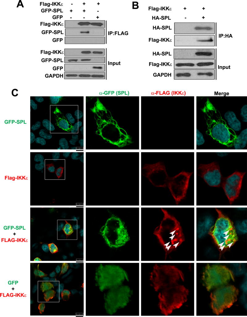
Figure 5. SPL interacts with IKKε.
(A) 293T cells were transfected with Flag-tagged IKKε (Flag-IKKε) in the presence of GFP-tagged SPL (GFP-SPL) or GFP expressing plasmid (GFP) or an empty control Flag vector (-). 24 hr after transfection, co-immunoprecipitation (IP) was carried out using anti-FLAG affinity resin and Western blotting analysis was performed to detect Flag-IKKε, GFP-SPL and GFP in the pull-down fractions (IP: FLAG) and in the whole cell lysates (Input). (B) 293T cells were transfected with Flag-IKKε in the presence of either an empty control vector (-) or HA-SPL. After 24 hr, co-IP was carried out using anti-HA coated affinity resin. Western blotting analysis was conducted to detect Flag-IKKε and HA-SPL in the pull-down fraction (IP: HA) as well as in the whole cell lysates (Input). (C) Flag-IKKε and GFP-SPL were either transfected alone or co-transfected together into 293T cells. Flag-IKKε and GFP expressing plasmid (GFP) were co-transfected into cells (GFP + Flag-IKKε) as a negative control. At 24 hr post-transfection, cells were stained with DRAQ5 to detect nuclei, which are shown in the merged images, and with antibodies against Flag (α-Flag) to detect IKKε and GFP (α-GFP) to detect SPL by confocal microscopy. Cells in the square boxes in the far left panels are shown in detail. White arrows indicate the punctate-like structure. Representative confocal images are shown. Scale bar represents 10 μm.

