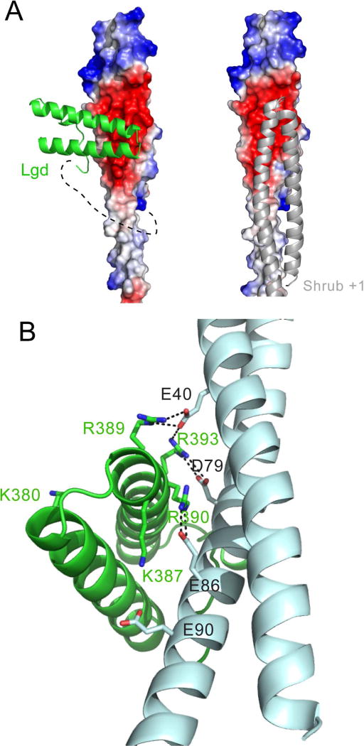
Figure 3.
Crystal structure of the Lgd-Shrub fusion protein. A, Left panel: Lgd-Shrub complex. DM14-3 is shown as a cartoon (green), and Shrub as an electrostatic surface on a sliding scale from negative (red) to positive (blue). The polyglycine linker connecting Lgd and Shrub, illustrated here with a dotted line, is not visible in the structure. Right panel: crystallographic model of a Shrub homopolymer (based on PDB entry 5J45). B, Key residues at the Lgd:Shrub binding interface. Lgd is shown as a cartoon in green. Shrub is shown as a cartoon in cyan. Charged residues at the interface are shown as sticks with salt bridges illustrated by dotted lines. See also Figure S3.

