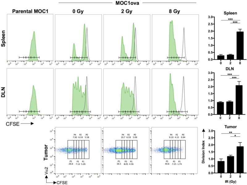
Figure 4. Antigen-specific T-lymphocyte priming in vivo following ionizing radiation.
Established (>0.1 cm3 volume) MOC1ova tumors in wild-type B6 mice were irradiated and CFSE-labelled OT-1 T-lymphocytes were adoptively transferred. CFSE spread of Vα2+ T-lymphocytes was assessed from the spleen, tumor-draining lymph node and tumor via flow cytometry (n=5 mice/group). Unfilled histograms represent CFSE-labelled OT-1 T-lymphocytes adoptively transferred into naive, non-tumor bearing mice. Representative CFSE histograms (spleen, lymph node) or dot plots (tumor) are shown with quantification of division index on the right. CFSE spread of adoptively transferred OT-1 T-lymphocytes into parental MOC1 tumor-bearing mice were used as a control for antigen-specific T-lymphocyte proliferation (left histograms). Representative results from one of at least two independent experiments are shown. *, p<0.05; **, p,0.01; ***, p<0.001.

