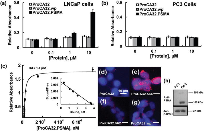
Fig. 4.

Characterization of PSMA targeting capacities of protein contrast agents at the cell level. (a, b) Comparison of the binding capability between ProCA32.wp and ProCA32.PSMA in LNCaP (a) and PC3 (b) cell lysates by ELISA. ProCA32.PSMA binds to LNCaP cell lysate at 1 and 10 μM. ProCA32.PSMA does not bind to PSMA-negative PC3 cells. (c) Determination of PSMA affinity by ELISA coupled with the Scatchard plot. (d–g) fluorescence staining of C4-2 cells incubated with ProCA32 (d), ProCA32.PSMA (e), ProCA32.562 (f), and ProCA32.wp (g). Among these PSMA-targeted ProCAs, ProCA32.PSMA shows the best PSMA targeting capacity. (h) Western blot shows the high expression of PSMA in C4-2 cells and no expression of PSMA in PC3 cells.
