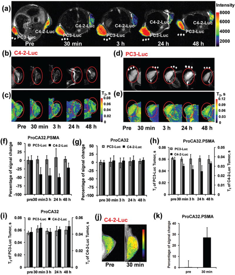
Fig. 6.

MR imaging of PSMA in xenografted mice tumors by protein based MRI contrast agents. (a) T2-weighted MRI of the mice implanted with both PC3-Luc and C4-2-Luc tumors before and after injection of ProCA32.PSMA. The PSMA-positive tumor, C4-2-Luc, exhibited decreased MRI signal intensity at 30 min–48 hours post injection of ProCA32.PSMA. After injection of ProCA32.PSMA, the PSMA-negative tumor, PC3-Luc, did not show any significant MRI signal change. (b) T2-weighted MRI of the C4-2-Luc tumor implanted mice before and after injection of ProCA32.PSMA. (c) T2 map of C4-2-Luc tumor implanted mice before and after injection of ProCA32.PSMA. (d) T2-weighted MRI of the PC3-Luc tumor implanted mice before and after injection of ProCA32.PSMA. (e) T2 map of PC3-Luc tumor implanted mice before and after injection of ProCA32.PSMA. (f) After injection of ProCA32.PSMA, the PSMA positive tumor (C4-2-Luc) shows a dramatically decreased signal between 30 min and 24 hours in T2-weighted MRI at 7 T. The PSMA negative tumor, PC-3-Luc, did not show any significant MRI signal change before and after injection of ProCA32. PSMA. (g) After injection of non-targeted ProCA32, the T2-weighted MRI signals in both PC3-Luc and C4-2-Luc were not changed due to the lack of PSMA targeting moieties in ProCA32. (h) After injection of ProCA32.PSMA, the C4-2-Luc tumor shows a decreased T2 value at 30 min, 3 hours and 24 hours. T2 of the C4-2-Luc returned to 33 ms at 48 hours post injection of ProCA32.PSMA. As a PSMA negative tumor, the T2 of PC-3-Luc did not have a significant signal change before and after injection of ProCA32.PSMA. (i) T2 of both C4-2-Luc and PC3-Luc tumors did not have significant change before and after injection of non-targeted ProCA32. (j) After I.V. injection of ProCA32.PSMA, the C4-2-Luc tumor in the xenografted mice showed increased MRI signal in the T1-weighted MRI. (k) The percentage signal increase plot of C4-2-Luc tumor in xenografted mice after injection of ProCA32.PSMA.
