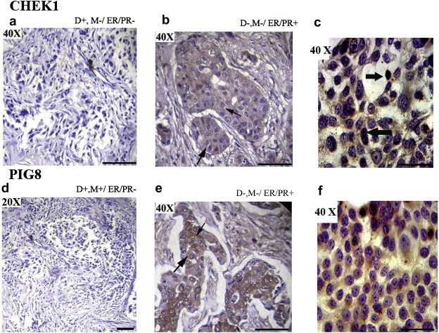
Figure 5.

Immunohistochemical analysis of CHEK1 and PIG8 in breast carcinoma samples and MCF‐7 cell line. (a) Breast carcinoma sample (#3156) showing reduced expression of CHEK1. (b) Breast carcinoma sample (#374) showing cytoplasmic expression of CHEK1. (c) MCF‐7 cell line showing nuclear expression of CHEK1. (d) Breast carcinoma sample (#5364) showing reduced expression of PIG8. (e) Breast carcinoma sample (#796) showing cytoplasmic expression of PIG8. (f) MCF‐7 cell line showing reduced cytoplasmic expression of PIG8. The slim arrow indicates cytoplasmic expression and bold arrow indicates nuclear expression. D: Deletion, M: Methylation, ER: Estrogen receptor, PR: Progesterone receptor. The original magnifications are indicated in top left corner of the photograph. Scale bar for magnification of 50 μM.
