
Figure 11.
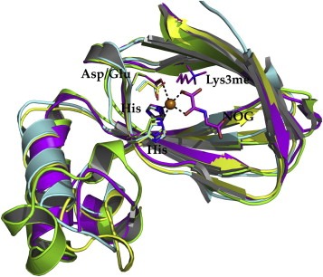
Superposition of KDM2A (yellow, PDB ID 2YU1), KDM4A (green, PDB ID 2WWJ), KDM6A (magenta, PDB ID 3AVR) and KDM7 (cyan, PDB ID 3U78) of the JmjC domain only (RMSD CA: 1.1Å). The residues chelating the iron ion are represented in sticks and color coded equivalent to their ribbon. The inhibitor N‐oxalylglycine (NOG) and the trimethylated lysine of 3AVR are shown in sticks and colored in magenta.