
Figure 5.
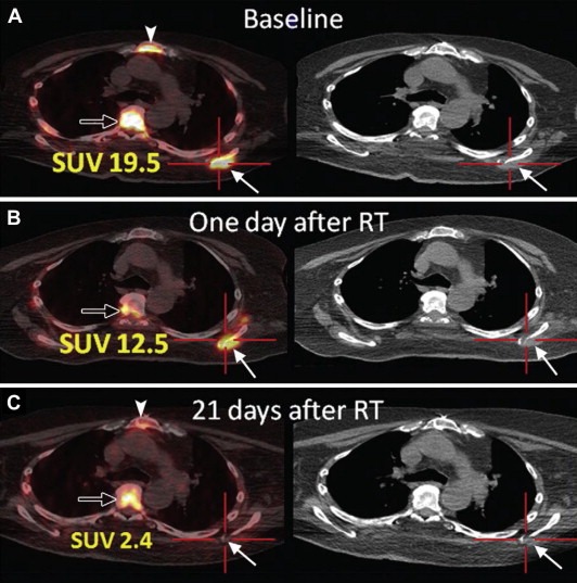
Images from PET/CT performed by using 18 F L‐thymidine (FLT) in a 70‐year‐old man with widely metastatic squamous cell cancer of the oropharynx. Images were acquired, A, before, B, 1 day after, and, C, 21 days after single dose fraction (2400 cGy) radiation treatment (RT) of a metastatic lesion in the left scapula (solid arrows). There is resolution of uptake at the tumor site in the left scapula. Physiologic FLT uptake is seen in bone marrow of the thoracic spine (open arrow) and sternum (arrowhead). SUV = standardized uptake value. (Reprinted from Hricak H. Oncologic imaging: a guiding hand of personalized cancer care. Radiology. 2011 Jun; 259(3):633–640).