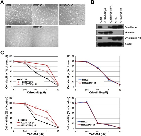
Figure 3.

TGF‐β1 stimulated EMT, resulting in resistance to ALK inhibitors. H2228 and H3122 cells were treated with TGF‐β1 (10 ng/mL) for 72 h, and these cells are referred to as H2228/TGF‐β1 and H3122/TGF‐β1, respectively. H2228/TGF‐β1 cells were incubated for 24 h in the complete medium without TGF‐β1, and these cells are referred to as H2228/TGF‐β1/R. (A) Cells were observed under a light microscope. (B) Cell lysates from each cell line were subjected to Western blot analysis. The indicated antibodies were used to evaluate EMT‐related marker proteins. (C) Cells were treated with the indicated doses of crizotinib or TAE‐684 in the presence or absence of TGF‐β1. Cell viability was measured using the MTT assay 72 h later. Bars represent the standard deviations.
