Figure 1.

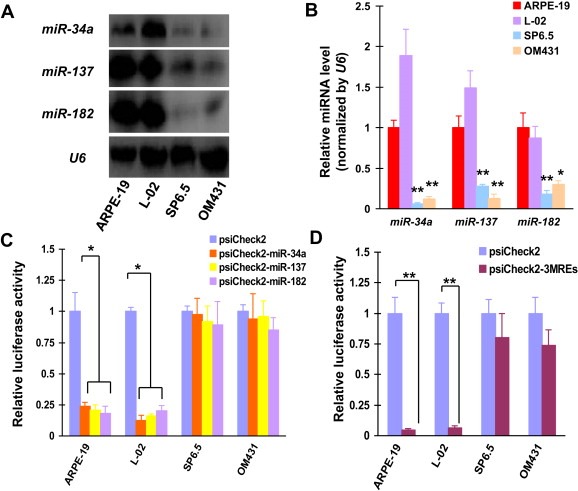
Uveal melanoma‐specific gene expression due to MREs of miR‐34a, miR‐137 and miR‐182. (A) Northern blotting was performed to detect the abundance of miR‐34a, miR‐137 and miR‐182 in ARPE‐19, L‐02, SP6.5 and OM431 cell lines. U6 was used as a loading control. (B) The levels of miR‐34a, miR‐137 and miR‐182 were also detected by qPCR in uveal melanoma and normal cell lines. Means ± SD of three independent experiments were shown. (C) Luciferase activity was detected 48 h after psiCheck2, psiCheck2‐miR‐34a, psiCheck2‐miR‐137 and psiCheck2‐miR‐182 transfection of normal and uveal melanoma cell lines. These experiments were performed in triplicate and means ± SD was presented. (D) Cells were also transfected with psiCheck2‐3MREs, followed by quantification of luciferase activity.
