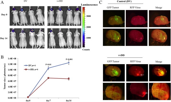
Figure 3.

vvDD inhibits the growth of intracranial AT/RT tumors in CD‐1 nude mice. BT16mGFPfLuc cells were stereotactically implanted into the brain of CD‐1 nude mice. After allowing two weeks for tumors to establish, mice were randomized into two groups (four animals each) that received a single intravenous dose of either live (vvDD) or killed vvDD (DV). The sizes of the tumors were quantified by the bioluminescent signals emitted from the tumors using the Xenogen IVIS 200 system as described in methods (A). The growth of the tumors was monitored at weekly intervals and plotted (B). Two weeks after treatment, the animals were sacrificed and photomicrographs of the brain tissues were taken to detect GFP‐expression (green, tumor) and mCherry signals (red, virus). Representative pictures are given (C).
