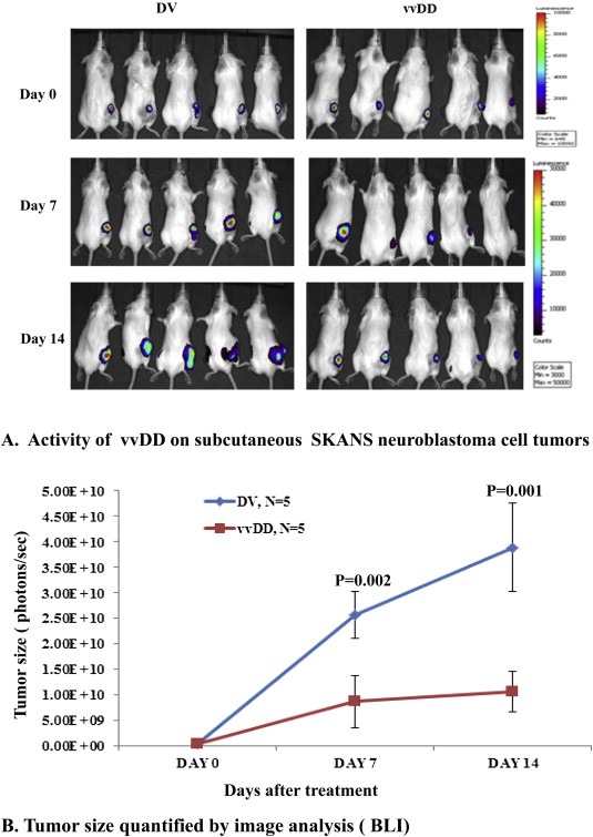
Figure 4.

In vivo activity of vvDD against neuroblastoma xenografts. CB17 SCID mice received SKNASmCherryfLuc neuroblastoma cells in the right flank to establish subcutaneous tumors. Bioluminescence imaging was carried out weekly to confirm tumor growth. Two weeks after tumor implantation, animals were assigned randomly into two groups to receive a single intravenous dose of 5 × 10⁷ PFU/mouse of vvDD or an equivalent dose of killed virus. Bioluminescent imaging was carried out at weekly intervals (A) and after approximately two weeks of receiving treatment, the animals were sacrificed and the size of tumor tissues was assessed (B).
