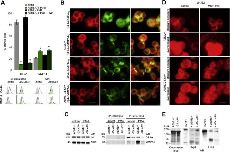
Figure 3.

The cooperativity of alpha6beta4 with MMP14 is impaired in ASML‐C4.4Akd cells: (A) ASMLwt and ASML‐C4.4Akd cells were stained with C4.4 and anti‐MMP14. Expression was evaluated by flow cytometry. Mean values ± SD (triplicates) of stained cells and representative examples are shown. Significant differences between ASMLwt and ASML‐C4.4Akd cells: *. (B) ASMLwt and ASML‐C4.4Akd cells were seeded on LN332 (804G supernatant)‐coated slides. Cells were double stained for C4.4A and alpha6beta4 or MMP14 or alpha6beta4 and MMP14. Single fluorescence staining and digital overlays are shown (scale bar: 10 μm). (C) ASMLwt and ASML‐C4.4Akd cells were seeded on LN332‐coated plates and were stimulated, where indicated, o/n with PMA (10−8M). Cells were lysed, precipitated with B5.5 (anti‐alpha6beta4) or control IgG and after SDS‐PAGE and transfer blotted with C4.4 and anti‐MMP14. WB of beta4 and actin are included as controls. (D) ASMLwt and ASML‐C4.4Akd cells were cultured overnight in the presence of PMA and DMSO (control) or MMP‐InhII on glass cover slides. Cells were stained with anti‐LN gamma2. The standard exposure and overexposure are shown. (E) PMA stimulated ASMLwt and ASML‐C4.4Akd cells were co‐cultured overnight with LN111 or LN332 (804G supernatant). Cells were removed by centrifugation. Supernatants were separated by SDS‐PAGE and stained with anti‐LN beta1 and anti‐LN gamma2. Staining of supernatants with Coomassie Blue is included as control. Co‐localization and co‐immunoprecipitation of alpha6beta4 and MMP14 is reduced in PMA‐stimulated ASML‐C4.4Akd cells. This is accompanied by a reduction in laminin degradation.
