Figure 5.

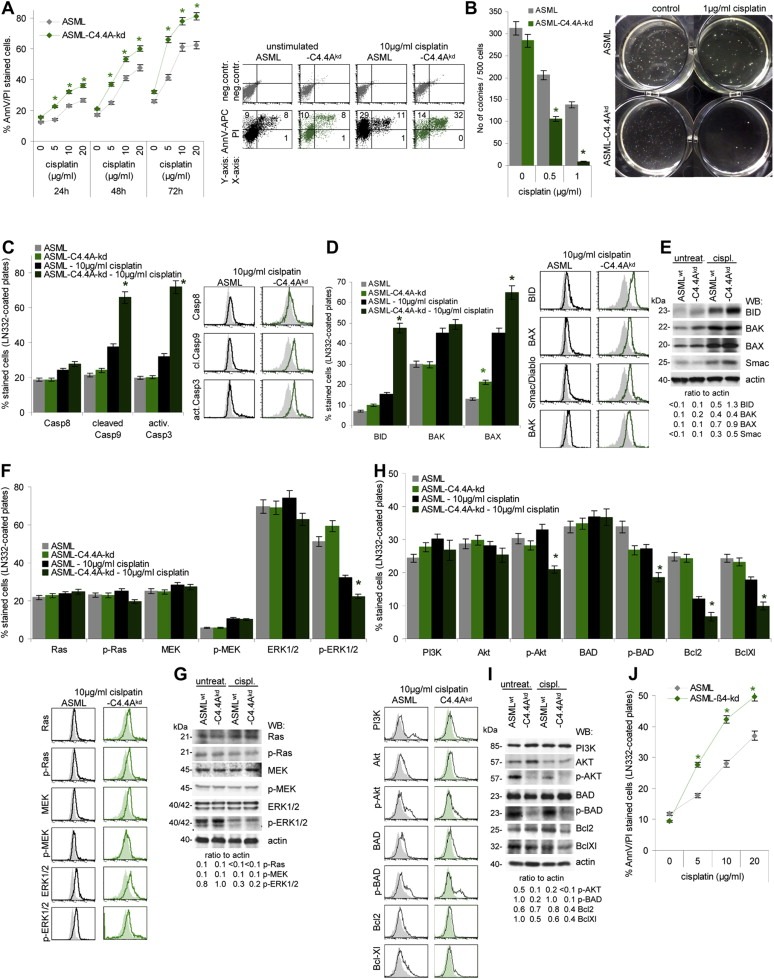
The contribution of the C4.4A‐alpha6beta4 cooperation to apoptosis resistance of ASML cells. (A,B) ASML and ASML‐C4.4Akd cells were cultured with titrated amounts of cisplatin for 48 h. (A) Apoptotic cells were determined by AnnV‐APC/PI staining after 48 h of culture (mean values ± SD of 3 experiments and representative example); (B) soft agar colony formation (mean valuesSD of 3 experiments and representative examples). In soft agar colony formation lower cisplatin concentrations were used, as growth in soft agar presents already a stress situation and cells are exposed to cisplatin for 3 weeks compared to 2 days in the apoptosis assays. Significant differences in cisplatin resistance between ASML and ASML‐C4.4Akd cells are indicated by *. (C‐I) ASML and ASML‐C4.4Akd cells, seeded on LN332, were cisplatin‐treated (10 μg/ml) for 48 h. Dead cells were discarded, performing the following analysis only with cells still alive. (C) Cells were stained with anti‐Casp8, anti‐cleaved Casp9 and anti‐activeCasp3; mean values ± SD of 3 experiments and representative examples are shown. (D) Cells were stained with anti‐BID, ‐BAK, ‐BAX, ‐Smac/Diablo; mean values ± SD of 3 experiments and representative examples are shown; (E) Lysates were SDS‐PAGE separated and blotted with the same antibodies as used in (D) and anti‐actin as control. The ratio of the signal intensity in comparison to actin is indicated. (F) Cells were stained with anti‐Ras, ‐p‐Ras, ‐MEK, ‐p‐MEK, ‐ERK1/2 and ‐p‐ERK1/2; mean values ± SD of 3 experiments and representative examples are shown. (G) Lysates were SDS‐PAGE separated and blotted with the same antibodies as in (F) and anti‐actin. The ratio of the signal intensity in comparison to actin is indicated. (H) Cells were stained with anti‐PI3K, ‐Akt, ‐p‐Akt, ‐BAD, ‐p‐BAD, ‐Bcl2, and ‐BclXl; mean values ± SD of 3 experiments and representative examples are shown. (I) Lysates were SDS‐PAGE separated and blotted with the same antibodies as in (H) and anti‐actin. The ratio of the signal intensity in comparison to actin is indicated. (A,B,C,D,F,H) Significant differences between untreated ASML and ASML‐C4.4Akd cells and between cisplatin‐treated ASML and ASML‐C4.4Akd cells: *. Repetition of the experiments with ASMLmock instead of ASMLwt and ASML‐C4.4Akd clone 30c instead of clone 34 cells revealed comparable results. (J) ASML and ASML‐ 4kd cells were seeded on LN332‐coated plates and cisplatin‐treated (10 μg/ml). Apoptosis was determined after 48 h by AnnexinV‐APC/PI staining. Significant differences between ASML and ASML‐ 4kd cells: *. ASML‐C4.4Akd cells show a significant decrease in drug resistance, most strongly in anchorage‐independent growth. Loss of drug resistance on LN332‐coated plates is not due to altered receptor‐mediated apoptosis, but proapoptotic molecules of the mitochondrial apoptosis pathway are upregulated. While activation of the MAPK pathway is hardly affected in cisplatin‐treated ASML‐C4.4Akd cells, phosphorylation of AKT and BAD is nearly abolished. As a transient beta4kd promotes a comparable loss in drug resistance, we hypothesize that C4.4A supports activation of the PI3K/Akt pathway via alpha6beta4 recruitment into rafts.
