
Figure 6.
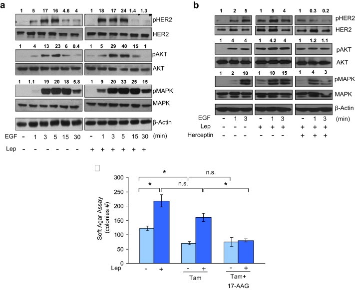
Effects of leptin on growth factor signaling and responsiveness to the antiestrogen tamoxifen. (a) MCF‐7 cells were treated with leptin 500 ng/ml (Lep) for 24 h and then treated with EGF 100 ng/ml as indicated. Levels of phosphorylated (p) HER2 (Tyr1248), Akt (Ser473), and MAPK (Thr202/Tyr204), at the indicated residues, and total non‐phosphorylated protein were measured in cellular extracts by immunoblot analysis. (b) MCF‐7 cells were treated with Lep for 24 h and then treated with EGF and herceptin (10 μg/ml) alone or in combination as indicated. Levels of phosphorylated (p) HER2 (Tyr1248), Akt (Ser473), and MAPK (Thr202/Tyr204), at the indicated residues, and total non‐phosphorylated protein were measured in cellular extracts by immunoblot analysis. β‐Actin was used as loading control. Numbers on top of the blots represent the average fold change versus untreated cells normalized for β‐actin. (c) Soft‐agar growth assay in MCF‐7 cells treated with Lep, tamoxifen (Tam, 1 μM) and 17‐AAG (20 nM) alone or in combination. After 14 days of growth, colonies >50 μm diameter were counted. n.s., nonsignificant; *p < 0.05.