
Figure 1.
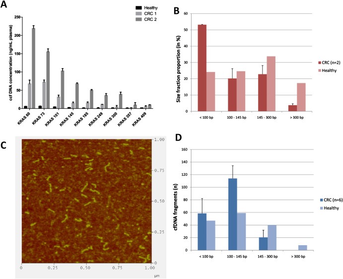
CcfDNA is highly fragmented. A. Size profile distribution of ccfDNA amounts in CRC (colorectal cancer) of two patients (named CRC1 and CRC2) and healthy individual plasma. The size profile distribution was evaluated with a Q‐PCR experiment and corresponding primer of targeting amplicons of increasing size. B. Proportions of ccfDNA per size range in % of the total observed ccfDNA with Q‐PCR. C. AFM visualization of ccfDNA fragments extracted and purified from stage IV CRC patient plasma. A representative visual determination of ccfDNA is shown here. D. Size profile distribution of ccfDNA fragments (n) measured with AFM experiments.