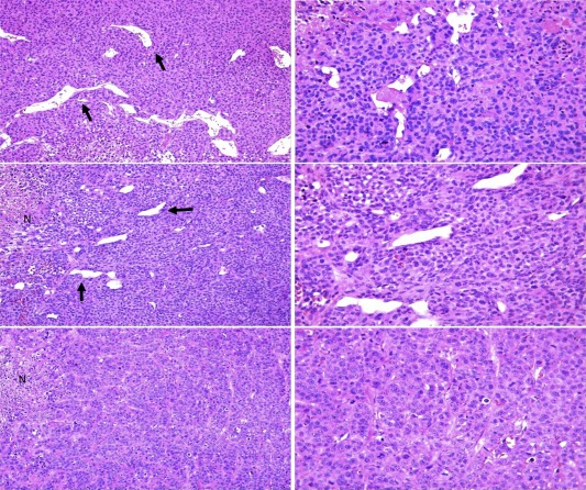
Figure 6.

In vivo vascular lacunae. Differential density of vascular lacunae (black arrows) in untreated (upper panels), Bevacizumab (middle panels) and Sunitinib (lower panels) treated engrafted tumors. While control cases showed the higher presence of vascular lacunae, their number progressively decreases in Bevacizumab treated mice. Sunitinib treatment exerted the strongest effects by inducing the decrease of vascular lacunae. Focal areas of necrosis (N) could be detected in both Bevacizumab‐ or Sunitinib treated mice. Original magnification ×100 left panels, ×200 right panels.
