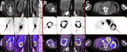
Figure 2.

Heterogeneous distribution of 124I‐girentuximab in RCC lesions. (A and B) Axial, sagittal, and coronal CT (top), PET (middle), and fused PET/CT (bottom) images of a patient with clear cell renal cancer with relatively homogeneous intratumoral distribution of the radiolabeled antibody (arrows) (A) and a patient with large, centrally necrotic clear cell renal cancer with marked heterogeneity (arrows) (B). (C) Axial CT (top), PET (middle), and fused PET/CT (bottom) images of a patient with advanced clear cell renal cancer. Antibody distribution within the primary tumor is heterogeneous (arrows), whereas distribution within metastatic nodes is relatively homogeneous (arrowheads). This figure was originally published in JNM; reprinted with permission (Pryma et al., 2011) (adapted).
