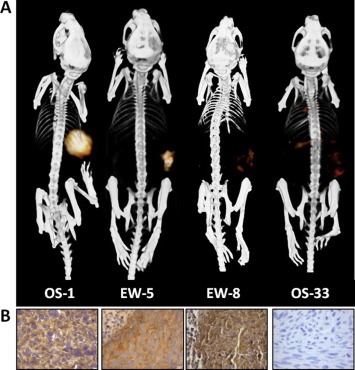
Figure 4.

111In‐R1507 immuno‐SPECT and immunohistochemical IGF‐1R expression in mice with subcutaneous bone sarcoma xenografts. 111In‐R1507 immuno‐SPECT/CT scans (A) and IGF‐1R expression levels of corresponding tumors (B) in mice with bone sarcoma xenografts. Xenografts with high (OS‐1), intermediate (EW‐5), or no (EW‐8 and OS‐33) response to treatment with the IGF‐1R antibody R1507 were selected. Images were acquired 3 days post injection of 111In‐R1507. This figure was originally published in Clin Cancer Res; reprinted with permission (Fleuren et al., 2011) (adapted).
