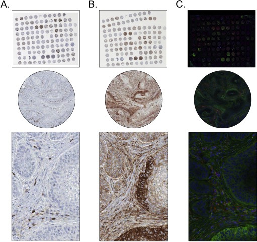
Figure 2.

A lung cancer TMA stained with antibodies towards PTPRC (DakoCytomation) and CD99 (DakoCytomation), utilizing both brightfield IHC (A and B) and darkfield IF (C) on consecutive sections. (A) and (B), IHC staining of PTPRC and CD99, respectively. PTPRC shows distinct cytoplasmic positivity in lymphoid cells, while CD99 is strongly expressed in both tumour cells and surrounding tumour stroma. The IHC stained images show clear tissue morphology and manual interpretation of staining intensity can be easily determined. (C), IF staining of PTPRC (red) and CD99 (green). The IF stained images show autofluorescence and not as clear morphology; however, the different dyes and antibodies can be easily distinguishable from each other.
