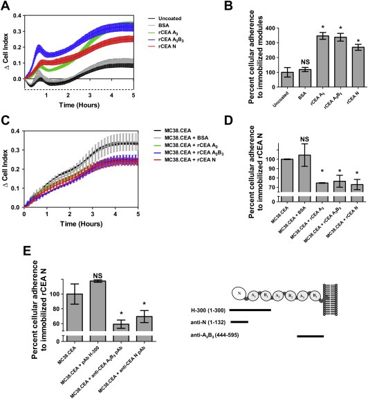
Figure 5.

Disruption of CEA‐homotypic binding impedes the adherence of CEA‐expressing tumor cells to rCEA N domain‐coated surfaces. A. Selective adherence of MC38.CEA cells to gold sensor E‐plates coated with either 1 μg BSA (control protein), rCEA N, A3 or A3B3 modules. Cellular adhesion of MC38.CEA cells to CEA modules was directly measured as changes in electrical impedance recorded by a gold sensor located in each tissue culture well of E‐plate wells. Changes in impedance, reported as ΔCell Index values, were measured at 1 min interval over a period of 6 h. B. Bar graph representing the percentage of cells adhering to the coated E‐plate surfaces at the end of the adherence phase of MC38.CEA cells (6 h post cell addition) relative to uncoated wells, set at 100% after “(6h post cell addition)”. Panels C and D highlight the disruption of cellular adherence to CEA N domain‐coated E‐plates in the presence of (1 μM) rCEA N, A3B3, or A3 modules, but not BSA, to MC38.CEA cells prior to dispensing these cells into wells. NS: not statistically significant; * significant when compared to the control protein (P ≤ 0.05). Statistical significance was established using a Student‐t‐test. E. Addition of polyclonal antibodies recognizing epitopes within regions of the N and A3 B3 domains of CEA suggest that antibodies directed at epitopes within regions 1–132 of CEA N domain (pAb H‐300) and residues 444–595 of the A3 B3 domain (anti‐CEA A3B3 pAb) can specifically inhibit CEA‐mediated homophilic cellular adhesion. NS: not statistically significant; * significant when compared to untreated MC38.CEA cells (P ≤ 0.05). Analyses were performed using a Mann‐Whitney‐U‐test.
