
Figure 3.
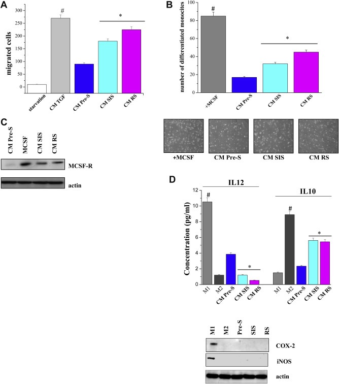
Conditioned media from senescent fibroblasts recruit human monocytes and differentiate them into M2 macrophages. A) Monocytes isolated from normal donor buffy coat were serum starved and then were allowed to migrate for 2 h toward CM of pre‐senescent or senescent fibroblasts. As positive control CM of fibroblasts activated with 10 ng/ml TGF‐β for 24 h was used. #p < 0.005 TGF‐β‐treated cells vs starvation; *p < 0.005 CM senescent cells vs CM pre‐senescent cells. B) Human monocytes were cultured for 7 days in CM from pre‐senescent or senescent fibroblasts. As a positive control of differentiation, monocytes were cultured for 7 days with MCSF (50 ng/ml). Differentiated macrophages were counted, and a bar graph, representative of six randomly chosen fields, is shown. #p < 0.005 MCSF treated cells vs untreated; *p < 0.005 CM senescent cells vs CM pre‐senescent cells. C) Monocytes were cultured as in B). Cells were lysed and the expression of MCSF‐R and actin was evaluated by immunoblots. D) M1 and M2 macrophages were obtained as reported in the Material and Methods section. Cells were treated as in B and the levels of IL‐12 or IL‐10 were measured by ELISA test. #p < 0.005 CM M1 cells vs CM M2 or CM fibroblasts; *p < 0.005 CM senescent cells vs CM pre‐senescent cells. Cell lysates were subject to immunoblot analysis to evaluate the expression of COX‐2, iNOS and actin.