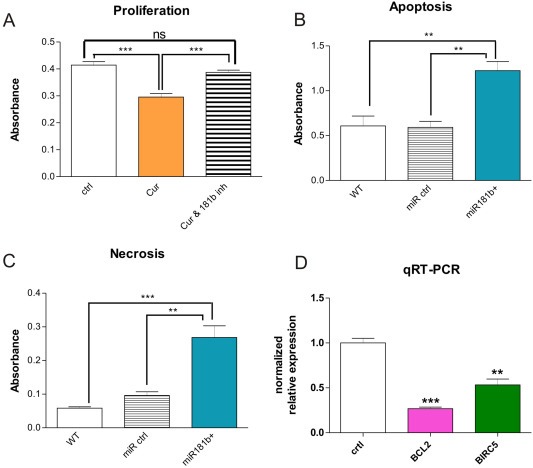
Figure 5.

Involvement of miR181b in tumor cell proliferation, apoptosis and invasion. A: Curcumin inhibits proliferation via miR181b. Treatment of MDA‐MB‐231 breast cancer cells for 24 h with Curcumin (“Cur”) inhibits cell proliferation. However, treatment with Curcumin together with a specific miR181b hairpin‐inhibitor (“Cur & 181b inh”) abolishes the anti‐proliferative effect of Curcumin, returning cell proliferation to control‐levels. The effect of miR181b expression on proliferation was statistically highly significant as evidenced by student's t‐test (***P < 0.001). "ns”: not significant. B, C: miR181b overexpression induces apoptosis and necrosis in breast cancer cells. Functional apoptosis/necrosis assays revealed that miR181b over‐expression led to enhanced apoptosis (left panel) as well as necrosis (middle panel). 24 h after transfection with miR181b oligos apoptosis rate in MDA‐MB‐231 cells was doubled as compared to wildtype MDA‐MB‐231 cells or to MDA‐MB‐231 cells transfected with an appropriate control oligo (left panel). Likewise, necrosis rate was increased approximately 5 fold in miR181b over‐expressing MDA‐MB‐231 cells as compared to wildtype cells and over 2.5 fold as compared to MDA‐MB‐231 cells expressing an appropriate control oligo (**P < 0.01; ***P < 0.001; ANOVA with Bonferroni's post test). Mean + SD from 3 independent experiments are shown. D: Using quantitative RT‐PCR, expression of the apoptosis related factors BCL2 and survivin/BIRC5 in MDA‐MB‐231 over‐expressing miR181b showed a statistically significant down‐regulation achieved 72 h after transfection with a double stranded miR181b oligo as compared to the appropriate controls (right panel) (**P < 0.01 and ***P < 0.001; student's t‐test). Mean + SD (SEM?) from 3 independent experiments are shown.
