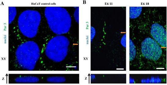
Figure 2.

Expression of high‐risk HPV‐18 E6 promotes the loss of Par3 protein from the apical domain of cell junctions. IF staining for Par3 (green) and nuclear DAPI staining (blue). Image reconstruction along the z‐axis was performed from Z stacks of mock‐transfected HaCaT cells (A, control) or derivative clones stably expressing HA‐E6 proteins (B) that were cultured on Matrigel for 72 h. In each case, a representative section from the x‐y axis is shown. The lower panel presents an individual x‐z section along the position indicated in the upper image (orange arrow). Scale bars: 5 μm.
