
Figure 4.
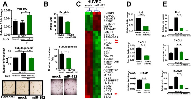
In vitro effects of ELV transfer in endothelial cells. A. Top: Quantification of miR‐192 assessed by qPCR in HUVEC previously incubated with 2 μg of ELVs from parental, mock, and miR‐192 cells for 72 h. Bottom: Tubulogenesis assay of HUVEC cells after 72 h of treatment with ELV isolated from parental, mock, and miR‐192 cells and representative images. B. Scratch and tubulogenesis assay of HUVEC cells after transfection with a pre‐miR‐192 or mock empty vector. C. Hierarchical cluster after integrative transcriptomic analysis of HUVEC cells overexpressing miR‐192 and mock transfected. Top list of the most significantly overexpressed (red) and repressed (green) genes are represented. Several angiogenesis‐related genes were found to be altered (red arrows). D. qRT‐PCR analysis confirmed the alteration of ICAM1, CXCL1, and IL‐8 in HUVEC cells transfected with miR‐192 or mock. E. qRT‐PCR analysis of HUVEC cells after 72 h incubation with 2 μg ELV isolated from parental, mock, and miR‐192 tumor cells.