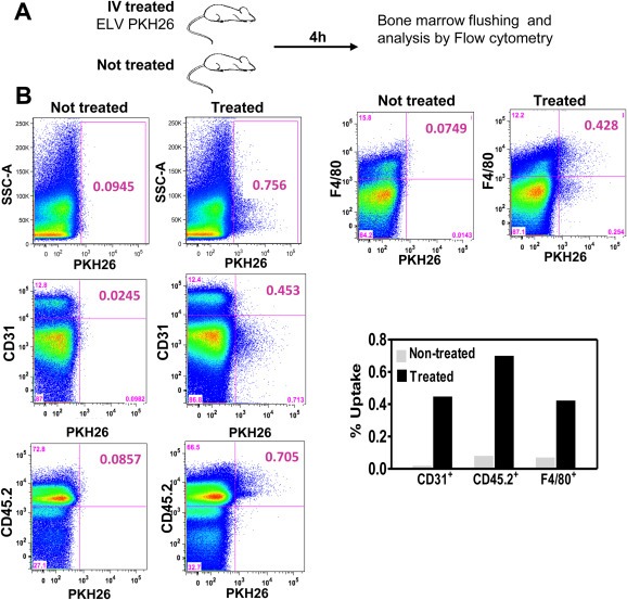
Figure 5.

In vivo transfer of ELV to cells in the bone marrrow compartment. A. Outline of the experimental setting. After PKH26‐labeling, fluorescently‐labeled ELV were injected and 4 h later, bone marrow flushing was performed and cells were analyzed by flow cytometry. Untreated mice were used as control. B. Bivariate displays of flow cytometry analysis showing an increase labeling of CD31+, CD45+ and F4/80+ cell populations in mice treated with PKH26–labeled ELV (right column) as compared to untreated mice (left column).
