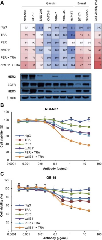
Figure 5.

oz1E11 shows antiproliferative activity in HER2‐overexpressing gastric and breast cancer cells A, Human gastric and breast cancer cells were treated with 5 μg/mL of antibodies for 3 or 4 days and cell viability was determined (upper panel). The expressions of HER2, EGFR, and HER3 in 20 μg of total cell lysate were determined by western blotting (bottom panel). Dose‐effect curves of antibodies in NCI‐N87 (B) and OE‐19 (C) cells are shown. Cell viability was expressed as the mean ± SD (n = 3), and the 100% point was defined as the absorbance of the untreated well.
