
Figure 3.
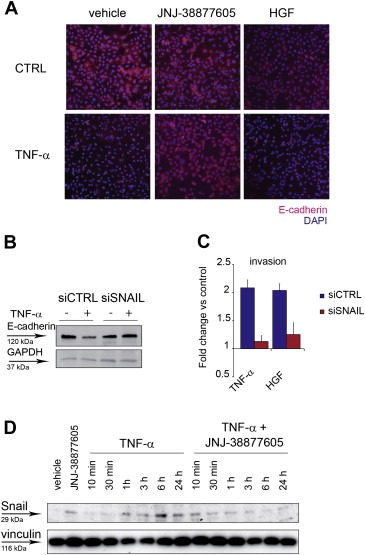
TNF‐α downregulates E‐cadherin via MET through Snail. (A) Immunofluorescence images (10×) of A549 cells treated with TNF‐α (10 ng/ml), in the presence of JNJ‐38877605 (500 nM), or vehicle (DMSO), or HGF (50 ng/ml), for 24 h. CTRL: cells without TNF‐α. Cells were stained with anti‐E‐cadherin antibodies (red). Nuclei were counterstained with DAPI (blue). (B) Western blot showing E‐cadherin protein in A549 transfected 48 h before with siRNA against Snail (siSNAIL) or control siRNA (siCTRL), and treated for 24 h with TNF‐α (10 ng/ml). GAPDH was probed as control of equal protein loading. (C) A549 invasion assessed in Transwell assay 24 h after treatment with TNF‐α (10 ng/ml), or HGF (50 ng/ml), in cells transfected 48 h before with siRNA against SNAIL (siSNAIL) or control siRNA (siCTRL). Graphs represent the fold change vs. control (untreated cells) of the number of invading cells. Bars: mean of three independent experiments ± S.E.M. (D) Western blot showing Snail protein in A549 cells at the indicated time‐points after treatment with TNF‐α (10 ng/ml), in the absence or in the presence of JNJ‐38877605 (500 nM). Vinculin was probed as control of equal protein loading.