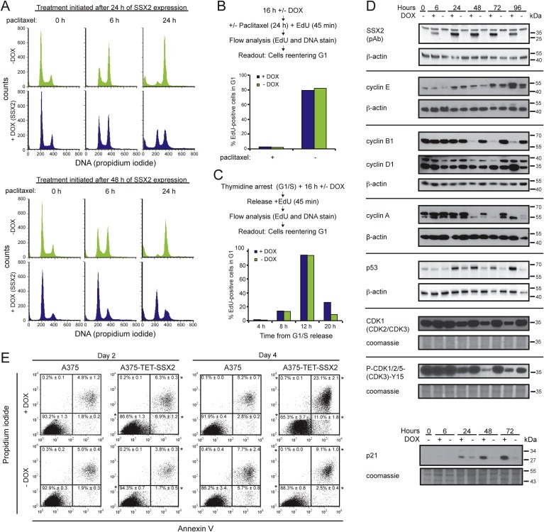
Figure 2.

SSX2 induces G1 checkpoint arrest and apoptosis in A375 melanoma cells. (A) Cell cycle analysis of A375‐TET‐SSX2 cells after paclitaxel treatment. Paclitaxel was added either 24 or 48 h after DOX induction of SSX2 and cells harvested after 0, 6 and 24 h of paclitaxel treatment. (B) Cell cycle kinetics. A375‐TET‐SSX2 cells with or without DOX‐induced SSX2 expression were treated with paclitaxel for spindle checkpoint activation and the number of cells reentering G1 was quantified by flow cytometry after release. Average of duplicates are shown. (C) Cell cycle kinetics. A375‐TET‐SSX2 cells, with or without DOX‐induced SSX2 expression, released from thymidine induced G1/S arrest, were EdU‐labeled and cells reentering G1 was quantified by flow cytometry. Average of duplicates are shown. (D) Western blot analysis of relevant cell cycle regulators in A375‐TET‐SSX2 cells with or without DOX‐induced SSX2 expression. (E) Apoptosis and dead cell analysis with Annexin V and propidium iodide. Each plot shows accumulated data from 3 independent samples, % ± standard deviation. *+DOX significantly differently from −DOX (p ≤ 0.01). The DOX concentration used in A–E was 50 ng/ml.
