
Figure 5.
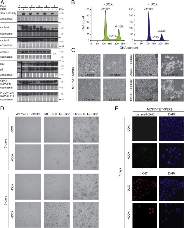
SSX2 induces a senescence‐like phenotype in MCF7 breast cancer cells. (A) Western blot analysis of relevant cell cycle regulators in MCF7‐TET‐SSX2 cells with or without DOX‐induced SSX2 expression. (B) Flow cytometric analysis of DNA content (Far Red Stain) shows an increase in the relative number of cells in G1 phase (48 h) in SSX2‐expressing MCF7‐TET‐SSX2 cells. (C) Long‐term growth of MCF7‐TET‐SSX2, A375‐TET‐SSX2 and HEK293‐TET‐SSX2 (H293‐TET‐SSX2) cells with SSX2 expression (10–14 days) induced an irregular and enlarged cells shape. DOX concentrations (induction of SSX2) are indicated. (D) Assay to detect senescence‐associated β‐galactosidase activity as represented by the ability of the cells to hydrolyze X‐gal and produce blue coloration. Cells were cultured for 3 or 6 days after induction of SSX2 expression before conducting the assay. (E) Immunocytochemical analysis of Ki67 and γ‐H2AX in MCF7 cells 7 days after induction of SSX2 expression. The DOX concentration used in A, B, D and E was 50 ng/ml.