Figure 3.

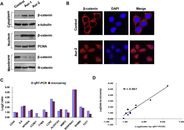
Overexpression of Aurora‐A alters subcellular localization of β‐catenin. A) The membranous, cytosol and nuclear proteins were isolated from the cells and analyzed with anti‐β‐catenin antibody. α‐tubulin, PCNA and N‐Cadherin were also examined respectively as the controls of different fractions. B) The subcellular distribution of β‐catenin was examined by immunofluorescence in control and Aur‐2 cells. The cells were fixed and stained with an anti‐β‐catenin antibody (red) and DAPI (blue). Pink color indicates overlap of red and blue colors. C) The upregulation of β‐catenin‐regulated genes by Aurora‐A. The levels of mRNA for β‐catenin‐regulated genes were analyzed using microarray and quantitative RT‐PCR in control and Aur‐2 cells. Fold inductions were obtained by normalizing mRNA levels of β‐catenin‐regulated genes in control cell line to that in Aur‐2 cells, and were shown by log2 ratio. D) Comparison of genes expression measurement by microarray and quantitative RT‐PCR. The expression changes in the selected 11‐β‐catenin‐regulated genes exhibited good agreement between the two experimental approaches.
