Figure 1.

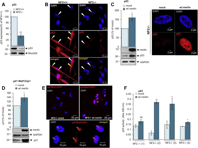
p53 levels and activity are decreased in schwannoma cells due to merlin deficiency. A. Western blot analysis demonstrates strong p53 expression in human primary Schwann (NF2+/+) and weak in human primary schwannoma (NF2−/−) cells. The levels of p53 are detected using anti‐p53 antibodies. ns (not significant): P > 0.05; *: P < 0.05; **: P < 0.01; ***: P < 0.001., mean ± SEM is given. B. Immunofluorescent staining of p53 (red) and DAPI (blue) in human primary Schwann (NF2+/+, left panel) and schwannoma (NF2−/−, right panel) cells. A stronger nuclear staining of p53 in Schwann (NF2+/+) compared to schwannoma (NF2−/−) cells. p53 strongly localises to the nucleus (white arrows) and weaker the cytosol (yellow arrows). C–F. Analysis of the effect of merlin reintroduction on p53 levels (C left panel), nuclear localisation (C right panel) and p53 activity [(D) p21 Waf1/Cip1 expression, (E) p21 Waf1/Cip1 cellular localisation and, (F) p53 transcription factor assay]. Cells were infected with mock virus or wild type (wt) merlin containing virus. ns (not significant): P > 0.05; *: P < 0.05; **: P < 0.01; ***: P < 0.001, mean ± SEM is given.
