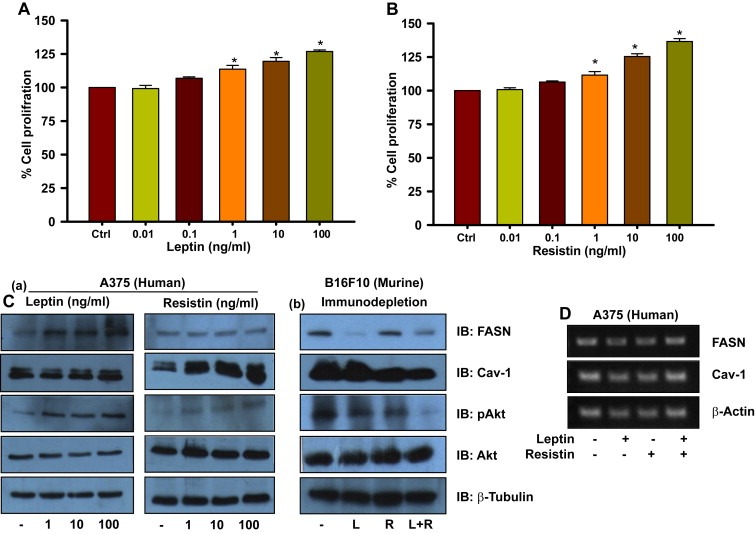
Figure 5.

Leptin and resistin promote proliferation, and modulate levels of FASN and Cav‐1 respectively, in melanoma cells. (A) MTT assay in leptin treated melanoma cells. A375 cells were plated in 96‐well plates. After 24 h, treatment with recombinant leptin was given at indicated concentrations in DMEM containing 1% FBS and cells were incubated for 48 h. (B) MTT assay for resistin treated melanoma cells. A375 cells were plated in 96‐well plates. After 24 h, treatment with recombinant resistin was given at indicated concentrations in DMEM containing 1% FBS and cells were incubated for 48 h. (C) Western blotting analysis of FASN, Cav‐1 and activated Akt. (a) A375 cells were plated in 35‐mm culture dishes. After 24 h, treatment with recombinant leptin or resistin was given at indicated concentrations in DMEM containing 1% FBS and cells were incubated for 48 h. These cell lysates were then subjected to SDS‐PAGE and Western blotting, (b) B16F10 cells were cultured in serum (collected from HFD C57BL/6J mice) which was immuno‐depleted of leptin and/or resistin for 48 h. Lysates of these cells were prepared and were subjected to SDS‐PAGE and Western blotting. (D) RT‐PCR analysis of 3T3‐L1 cells treated with leptin and/or resistin. A375 cells were treated with leptin and/or resistin (100 ng/ml) for 48 h as described in Materials and methods. The results are given as means ± standard deviation; *, p < 0.05; L, leptin; R, resistin.
