
Figure 5.
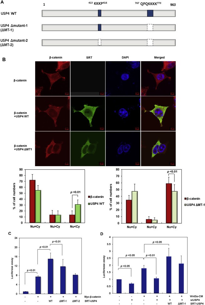
The role of nuclear localization sequence (NLS) in USP4. (A) Construction of the USP4 mutants lacking the NLS sequence (ΔMT‐1 and ΔMT‐2). (B) Subcellular localization of ΔMT‐1. After transfection of wild‐type USP4 or ΔMT‐1 along with Myc‐β‐catenin, the location of each protein was examined by immunofluorescence using anti‐SRT (green) and anti‐β‐catenin (red) antibodies, respectively (Top). Nuclei were stained with DAPI (blue). The cellular localization of the expressed proteins was examined in 100 immunostained cells and their relative abundance is represented by percentage bar graphs (bottom) using the same criteria in Figure 3C. (C) The effect of the NLS deletion mutants on the TOP flash reporter activity in the presence of WNT signal. The plasmids encoding wild‐type USP4, ΔMT‐1 and ΔMT‐2 were transfected into 293T cells along with Myc‐β‐catenin. After 48 h, TOP flash activity was measured. (D) The effect of NLS deletion mutant of USP4 on the TOP flash reporter activity in siUSP4‐treated HCT116 cells. HCT116 cells were transfected with siUSP4 for 6 h, followed by complementation with the wild‐type (WT) or ΔMT‐1 USP4. After 24 h, the cells were cultured in control‐CM or Wnt3a‐CM for an additional 24 h before the reporter assay.