
Figure 6.
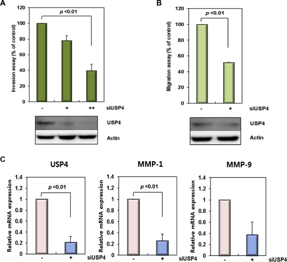
USP4 is correlated with β‐catenin in colon cancers. (A) Invasion activity of siUSP4‐treated colon cancer cells. HCT116 cells were treated with USP4 siRNA (siUSP4; +: 40 pM, ++: 90 pM) for 24 h, and in vitro cell invasion assays were performed for 48 h. (B) Cell migration ability of siUSP4‐treated colon cancer cells. HCT116 cells were treated with 90 pM siUSP4 for 48 h and assayed for their migration ability for 20 h. The invasion and migration activities of the siUSP4 transfected cells were determined relative to those of scrambled siRNA‐treated cells. The expression level of USP4 was analyzed by immunoblotting. Data shown represent the average values from three independent experiments. (C) Expression of migration and invasion markers in the siUSP4‐treated colon cancer cells. The expression level of USP4, MMP‐1, and MMP‐9 was examined 24 h after siUSP4 treatment in HCT116 cells. The mRNA levels were quantified by real time‐PCR with normalization to GAPDH. (D) The correlation between the expression levels of USP4 and β‐catenin, and Cyclin D1 and β‐catenin in colon cancer tissues. The mRNA levels of USP4, β‐catenin, and Cyclin D1 were measured by quantitative real time‐PCR according to an internal calibrator using the 2−△△CT method with normalization to GAPDH. This experiment was repeated twice. (E) Expression of USP4 and β‐catenin in colon cancer tissues. The levels of USP4 and β‐catenin proteins in colon cancer and corresponding normal tissues were measured by immunoblotting using GAPDH as a loading control. T, colon cancer samples; N, normal samples. (F) Expression of β‐catenin (a, c, e) and USP4 proteins (b, d, f) in normal tissues (a, b), primary (c, d), and metastatic colon cancers (e, f) (top). The representative regions of each sample (red boxes, top a–f) are magnified (bottom, a′–f′). In normal colon tissues, β‐catenin (a and a′) and USP4 (b and b′) showed expression in mucosa. Staining of colon adenocarcinoma revealed high levels of β‐catenin (c and c′) and USP4 (d and d′). β‐catenin (e and e′) and USP4 (f and f′) were also actively expressed in metastatic carcinoma that had metastasized from the colon to the ovary. Both adenocarcinoma and metastatic carcinoma tissues were taken from the same patient.