
Figure 1.
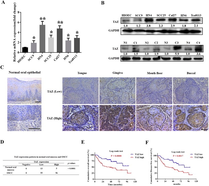
TAZ is overexpressed in OSCC and associates with patients survival A: TAZ mRNA levels were measured by real‐time RT‐PCR in six OSCC cell lines as compared with human immortalized oral epithelial (HIOEC). B: TAZ protein levels were determined by western blot (WB) in OSCC cell lines and paired tumor‐adjacent non‐tumor tissues (n = 4). Representative images of WB are shown. The “N” stands for non‐tumor tissue, and “C” stands for cancer in the lower panel. C: TAZ expression in human normal oral mucosa and OSCC specimens from diverse primary sites was evaluated by immunohistochemical staining. Rare TAZ positive staining was observed in basal cells from healthy oral epithelial. Scale bar: 100 μm. D: Expression patterns of TAZ in human OSCC samples and normal counterparts were statistically determined. E, F: Overall and disease‐free survival analyses of patients with high or low expression of TAZ were estimated by Kaplan–Meier method and compared with log‐rank test. Data shown here are mean ± SD from three independent experiments. *p < 0.05, **p < 0.01, ANOVA analysis.