
Figure 3.
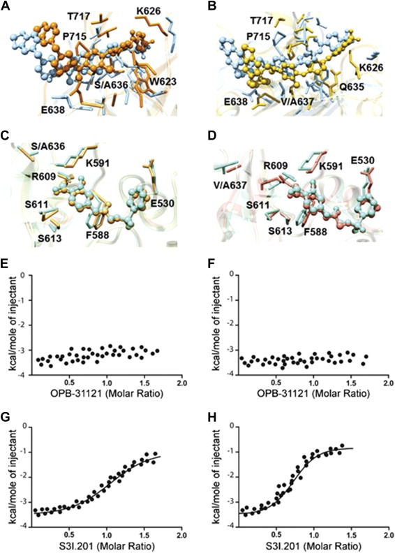
Mutational analysis of OPB‐31121 binding site in the STAT3 SH2 domain. (A) Superposition of the binding site of wild type (light blue) and S636A STAT3 mutant (orange) in complex with OPB‐31221. (B) Superposition of the binding site of wild type (light blue) and V637A STAT3 mutant (golden rod) in complex with OPB‐31221. (C) Superposition of the binding site of wild type (aquamarine) and S636A STAT3 mutant (sandy brown) in complex with S3I.201. (D) Superposition of the binding site of wild type (aquamarine) and V637A STAT3 mutant (salmon) in complex with S3I.201. In all panels drugs are depicted as colored sticks‐and‐balls, while main residues involved in the interactions are labeled and shown as colored sticks. Hydrogen atoms, water molecules, ions and counterions are omitted for clarity. (E) ITC data for S636A mutant STAT3 SH2 domain in complex with OPB‐31121. (F) ITC data for V637A mutant STAT3 SH2 domain in complex with OPB‐31121; (G) ITC data for S636A mutant STAT3 SH2 domain in complex with S3I.201; (H) ITC data for V637A mutant STAT3 SH2 domain in complex with S3I.201.