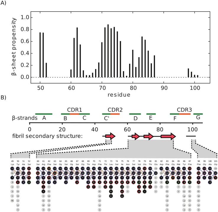
Fig 2. Secondary structure analysis of MAK33 VL in the fibril state.
A) β-sheet propensity calculated with TALOS+ [32]. B) Sequence and secondary structure elements of the native VL fold. Green and red bars indicate β-strands and CDRs of the native structure, respectively. Red arrows below the sequence indicate β-strands in the fibril state. The expansion shows the assigned atoms in the aggregated state.

