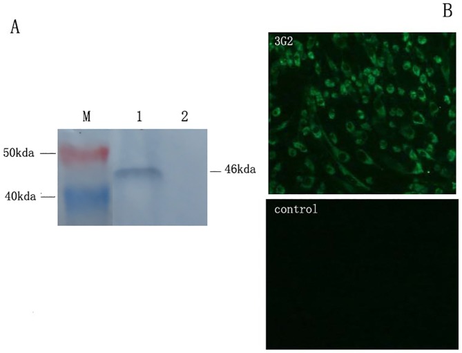
Fig 2. IFA and western-blot identification of mAb 3G2.
A: Western-blot identification Lane 1, Control, GST-tag didn’t react with mAb 3G2; Lane 2, The band of NS1-GST fusion protein was reacted with mAb 3G2; Lane M, Blue plusIIprotein Marker (14-120kda, Transgen Biotech). B: IFA identification Monoclonal antibody against TMUV NS1 protein was used to perform IFA on TMUV-infected BHK-21 cells. BHK-21 cells infected with TMUV yielded significant fluorescence with six MAbs in the cytoplasm; Control BHK-21 cells didn’t yield any fluorescence.

